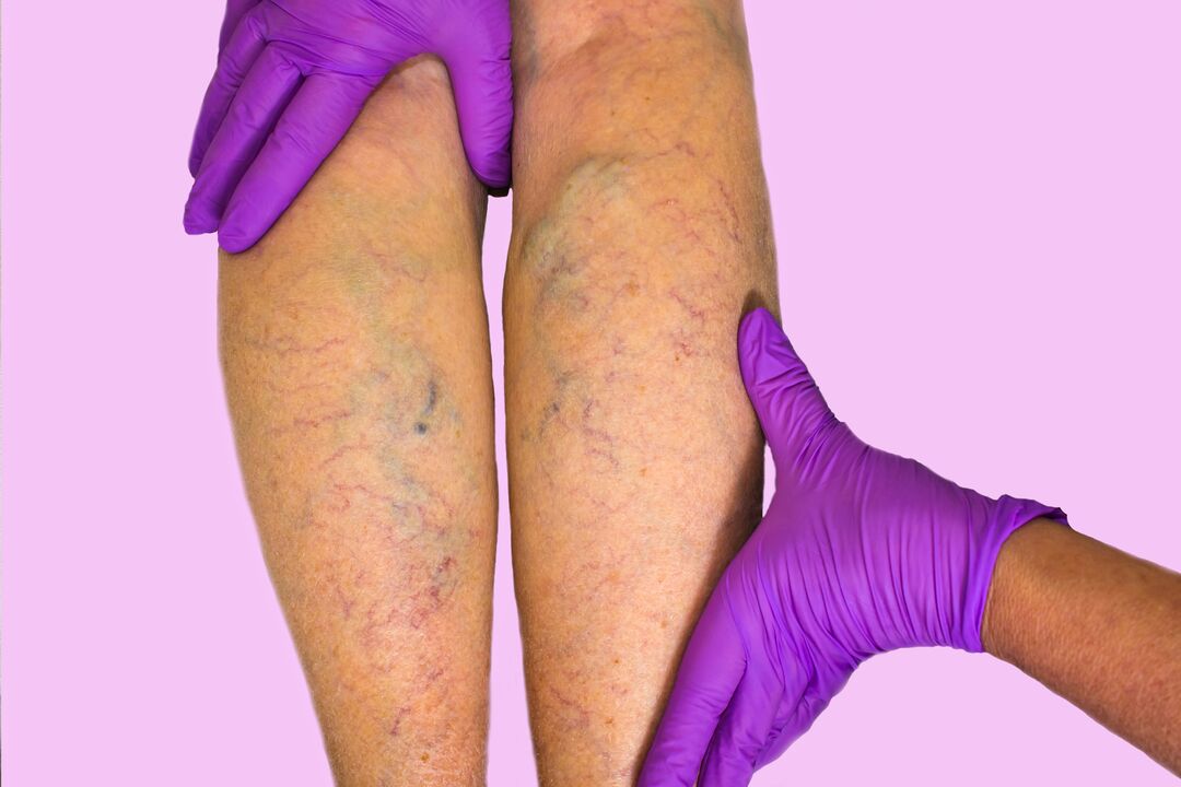
Varicose veins are pathological expansion of the lumen of the veins, which is caused by the thinning of their walls and a decrease in their tone. In advanced stages, venous nodes bulge under the skin and can occasionally become inflamed. Varicose veins are not just an aesthetic defect. The symptom refers to a blood circulation disorder, which impairs the quality of nutrition of tissues and organs and increases the risk of blood clot formation. Early diagnosis and treatment can slow down the development of pathology and prevent complications.
About the disease
Varicose veins are a chronic disease that includes any disturbance in the structure and function of the venous system. These can be congenital, genetically determined disorders, as well as pathological changes occurring as a result of external factors.
Varicose veins affect about 60% of adults worldwide, mainly Europeans. Most often, women suffer from varicose veins. The reason for this is the presence of a relationship between the tone of the vascular walls and the hormonal level.
Types of varicose veins
Varicose veins usually mean the enlargement of the veins of the legs, but pathological changes can also affect other parts of the body. Depending on the location, there are:
- rectal varicose veins (golden veins);
- dilation of the veins of the esophagus;
- varicose veins of the spermatic cord in men (varicocele);
- varicose veins of lower limbs.
Sometimes reticular varicose veins are isolated separately. It consists of blood vessels and stars visible under the skin. It mainly occurs on the legs, but it can appear under the breasts, on the abdomen and on other parts of the body. The disease is diagnosed when the saphenous veins of the reticular bed are dilated in the reticular layer of the dermis. It occurs in 50% of women. The formation of nodes is not typical.
Types of varicose veins of the limbs according to the CEAP classification (stages of development):
- C0 – no signal;
- C1 – appearance of varicose veins and stars;
- C2 – varicose veins;
- C3 – swelling of the legs appears, indicating the development of venous insufficiency;
- C4 – trophic changes in the form of hyperpigmentation, lipodermatosclerosis (thickening of the skin of the lower third of the leg);
- C5 – healing venous ulcers;
- C6 – non-healing venous ulcers.
Symptoms
The main symptoms of varicose veins of the lower limbs are as follows:
- heaviness in the legs (calves), swelling, worse in the evening;
- increased leg fatigue;
- aching pain in the calf that occurs in a long static position, standing or sitting.
With the development of the pathology, blue, tortuous veins begin to bulge under the skin, sometimes swelling to the point of knots. A sign of chronic venous insufficiency is a change in skin color, which is associated with damage to tissue trophism (nutrition). Extensive eczematous redness, itchy blisters and nodules appear. The swelling of the legs does not go away even after a night's rest.
Signs of the reticular form of varicose veins are limited to the subcutaneous vasculature. There may be heaviness in the calf and slight itching in the area of the dilated blood vessels. Trophic abnormalities are usually not observed.
Causes of varicose veins
Reticular varicose veins are caused by the replacement of collagen type 1 in the walls of blood vessels with collagen type 3. As a result, their contractility deteriorates - after dilation, the blood vessels no longer return to their original state. The cause of vessel wall thinning is the excessive activity of enzymes that destroy extracellular matrix proteins and elastin.
In women, the hormone progesterone helps to reduce the tone of the smooth muscle fibers of the vessel walls. Estrogen reduces the level of antithrombin, increases blood coagulation and provokes the development of stagnant processes.
The main cause of varicose veins of the limbs, accompanied by the appearance of lumps and bumps, is the failure of the valve mechanisms. Valves are folds formed by the inner lining of the veins. Normally, they work in only one direction: they open under the pressure of blood flow and do not let it back. If the valve mechanism is weakened, the blood flows back (reflux), which causes the walls of the veins to expand and the inner lining to become inflamed. Then the pathological process spreads to the deeper layers of the venous wall. His muscle fibers begin to be replaced by scar tissue, atrophy occurs. The walls no longer contract and their collagen structure is destroyed. The vein twists in a spiral.
The increased pressure in the vessels causes the failure of the musculo-venous pump. This is a system that controls the pumping of blood to the muscles during exercise and relaxation ("peripheral heart"). The result is congestion and venous insufficiency.
The provoking factors are:
- heredity: in most cases, varicose veins are caused by mutations in genes responsible for the structure of venous valves and walls;
- overweight;
- sedentary lifestyle;
- increased load on the venous system of the limbs due to standing work;
- pregnancy and childbirth, menopause, hormonal imbalance.
Varicose veins can be caused by poor foot movement due to uncomfortable shoes, as well as bad habits: smoking, alcohol consumption.
Diagnostics
The main methods of diagnosing varicose veins include a visual examination by a vascular surgeon, during which he assesses the condition of superficial and deep veins and identifies signs of tissue malnutrition. The patient is then sent for further diagnostics.
- Ultrasonic duplex scanning. It allows assessing the condition of the valves, the strength and direction of blood flow, the size of blood vessels, and identifying the presence of blood clots.
- Study of valve functions: compression tests, simulated walking, Parana maneuver.
- X-ray contrast venography is an X-ray in which contrast material is injected into the veins. It helps to assess valve function, vein patency and detect blood clots.
To clarify the diagnosis, the doctor may prescribe CT, MRI, thermography, radiophlebography, intravascular ultrasound, coagulation blood tests, etc.
Expert opinion
Varicose veins are not just unsightly veins that protrude under the skin. The complications of varicose veins are extremely unpleasant.
- Trophic disorders. Large brown spots appear on the legs or thighs and later develop into large, non-healing ulcers that are itchy and painful.
- Thrombophlebitis is inflammation of the vein walls, accompanied by the deposition of thrombotic masses. The thrombosed vein reddens, thickens, hurts, and the temperature rises around it. It looks like an abscess. It threatens the spread of infection in the body.
- Bleeding. Bleeding from a ruptured varicose vein can occur both inside and outside the tissue. The bleeding is intense and an ambulance should be called.
- Thromboembolism. A blood clot in an enlarged vein can break off and block vital arteries, such as the pulmonary artery. This condition often leads to instant death.
Timely diagnosis helps prevent serious consequences of varicose veins and identify the causes that provoked them.
Treatment of varicose veins
The specific treatment of reticular varicose veins involves several areas.
- Compression therapy - Wearing Class A and Class I supportive knitwear (socks, stockings) to prevent backflow of blood.
- Pharmacotherapy - taking phlebotonic drugs to increase the tone of the veins. These remedies do not remove the external signs, but they eliminate subjective symptoms such as heaviness, swelling and aching pain.
- Phlebosclerosis is the gluing of small blood vessels by the introduction of sclerosing substances. Microsclerotherapy allows you to get rid of vascular networks.
- Laser therapy - enables the elimination of minor defects remaining after microsclerotherapy. During the procedure, the doctor applies a beam of light to the affected areas.
An important part of the therapy is exercise, weight loss, wearing comfortable shoes and regular physical activity.
For the surgical treatment of varicose veins, together with the appearance of nodes, two methods are used: classic phlebectomy and endovenous thermal obliteration. The first method is considered obsolete. It involves ligating the junction of the vein with the common femur and removing the affected part of the trunk. The method is characterized by increased trauma and a high risk of relapse.
Thermal obliteration is a gentle, minimally invasive treatment method. The doctor inserts a laser catheter into the vein through a small incision, turns on the radiation, and slowly withdraws it. As the laser moves, it seals the vein by increasing the temperature. After that, it gradually resolves.
Prevention
In order to prevent the development or recurrence of varicose veins, persons at risk should:
- minimizes the static load on the legs;
- eat sensibly and, if necessary, take venotonic as prescribed by the doctor;
- wear compression stockings when standing in a static position for long periods of time.
It is useful if you regularly do cardio exercises to train the heart and blood vessels.
Rehabilitation
During the recovery period after surgery, the patient should wear compression stockings, reduce the load on the legs to a minimum, avoid overheating, and take medications prescribed by the doctor. The total rehabilitation time depends on the extent of the intervention and the presence of complications.


